X-Rays (Oxford AQA IGCSE Physics)
Revision Note

Author
Leander OatesExpertise
Physics
Properties of X-Rays
X-rays are part of the electromagnetic spectrum
X-rays have:
a very short wavelength
a very high frequency
high energy
X-rays are ionising radiation
They can remove electrons from atoms
This means that they can damage the structure of DNA and therefore cause damage to cells
The properties of X-rays include:
They affect photographic film in the same way as light
They are absorbed strongly by metal and bone
they are transmitted by healthy tissue
These properties make X-rays very useful for medical imaging
Uses of X-Rays
X-rays are used in medicine for both diagnostics and treatments
X-ray imaging
X-ray images are used to diagnose medical and dental issues
X-rays are directed at the patient
The X-rays are absorbed by bone or teeth, but pass through tissue
The X-rays that pass through the patient are detected by a flat-panel detector or photographic film
An image is created showing the bones or teeth of the patient
Contrast mediums can be used to view organs using X-rays
A contrast medium is a substance that absorbs X-rays, for example, barium
The patient is given food or drink containing the contrast medium (for viewing the digestive or renal system)
Or the patient is injected with the contrast medium (for viewing arteries or blood vessels)
An X-ray image is conducted
The organ or structure containing the contrast medium will absorb the X-rays and can therefore be shown on the image
X-ray using barium as a contrast medium

CT scans
X-rays are used in computerised tomography scanners (CT scanners) to create cross-sectional images through the body and three-dimensional (3D) images of organs
CT scan
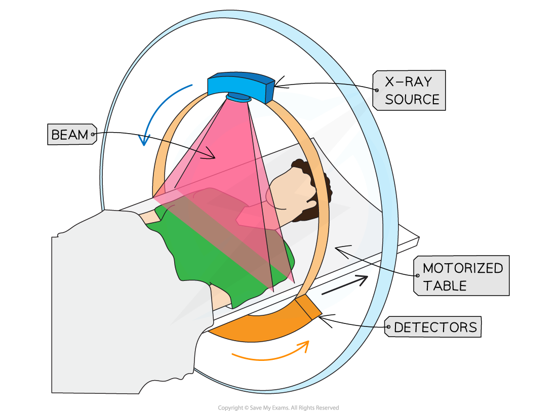
The X-ray tube moves around the ring
The X-rays pass through the patient
The X-rays are detected by the ring of detectors
The computer produces an image of the scanned area
The CT scan can distinguish between different types of soft tissue
X-ray therapy
X-rays are also used to treat certain conditions
Such as killing cancer cells
Shorter wavelength X-rays are used for treatment
These have a higher frequency and a higher energy
Metal filters are used direct the correct wavelengths to the correct location
A thin sheet of metal is placed between the X-ray tube and the patient
The metal filter absorbs the X-rays at other wavelengths
This minimises the exposure to the patient's healthy tissues

You've read 0 of your 0 free revision notes
Get unlimited access
to absolutely everything:
- Downloadable PDFs
- Unlimited Revision Notes
- Topic Questions
- Past Papers
- Model Answers
- Videos (Maths and Science)
Did this page help you?