Structure of the Eye (Oxford AQA IGCSE Physics)
Revision Note

Author
Ann HowellExpertise
Physics Content Creator
Structure of the Eye
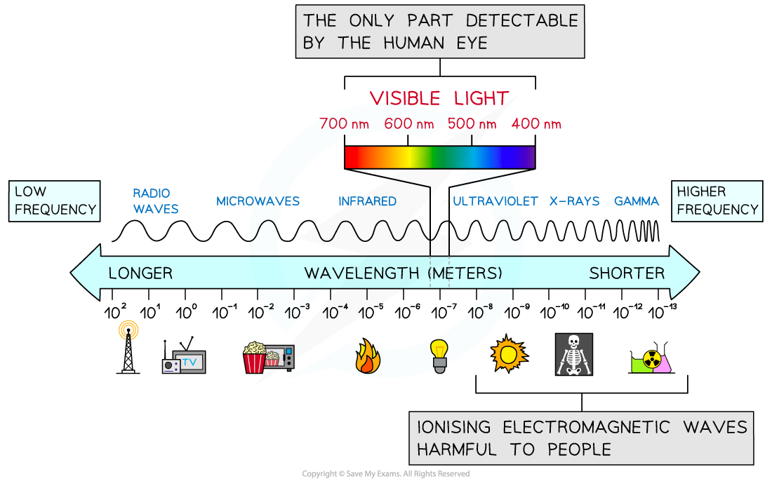
Our eyes only detect a limited range of electromagnetic waves known as visible light
Visible light is made up of wavelengths that form the colours of the spectrum:
Red
Orange
Yellow
Green
Blue
Indigo
Violet
The electromagnetic spectrum
The structure of the eye
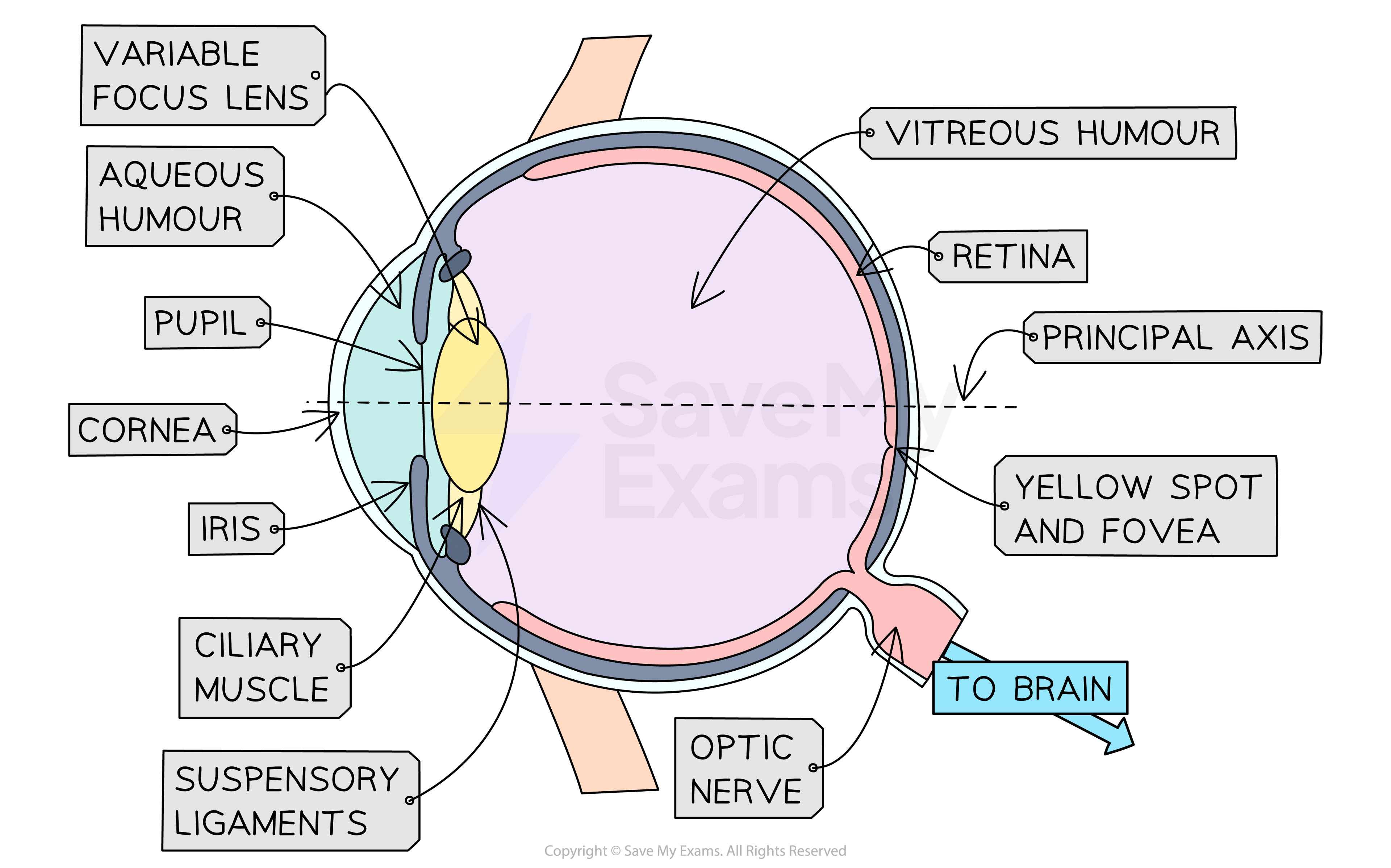
The cornea
Light rays enter the eye through the cornea
The cornea is a transparent layer that covers the front of the eye to protect it
It helps to focus light on the retina
When passing through the cornea light rays are refracted (change direction)
The lens and the ciliary muscles
The lens focuses light onto the retina
Light is also refracted as it passes through the lens
The lens is referred to as a variable focus lens because it can change its shape to focus on objects at different distances away
The lens is controlled by the ciliary muscles which are attached to the lens by suspensory ligaments
The muscles are attached to fibres which pull and stretch the lens
This changes the thickness of the lens
Which controls the eye's focal length
When the muscles contract the lens becomes thicker and more powerful
This occurs when the eye is focusing on an object close by
When the muscles relax the lens becomes thinner and less powerful
It occurs when the eye is focusing on an object close to the far point
The retina
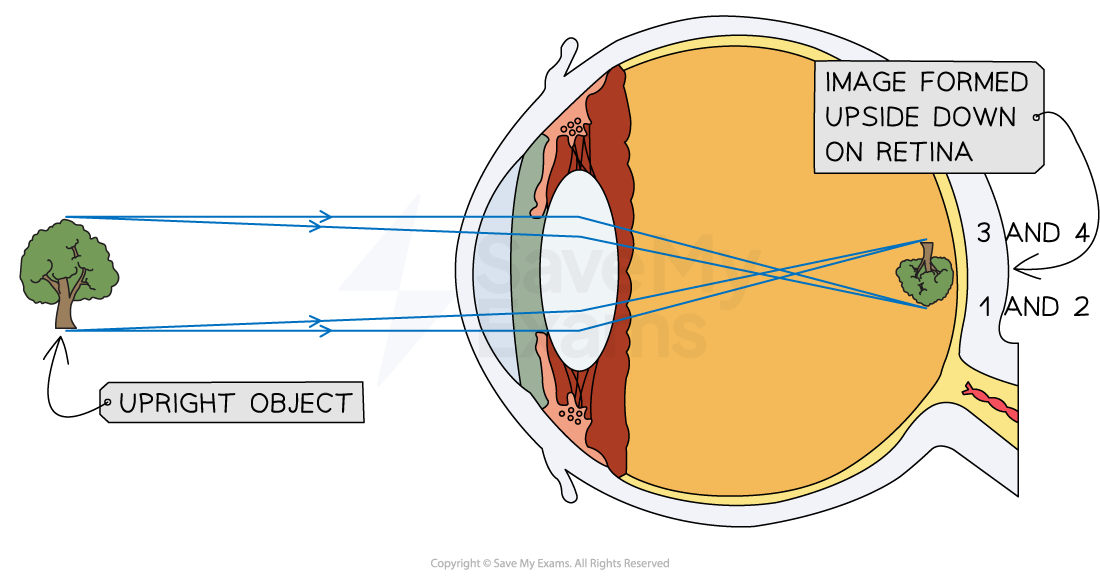
After passing through the lens the light is focused on the retina
The retina is made up of light-sensitive cells around the inside of the eye
The light rays are refracted through the cornea and lens so that they cross within the eye and arrive at the retina the opposite way around
Rays from the top of the object are now at the bottom of the retina and vice versa
The brain interprets the image so it is the correct way up
The inverted image on the retina
The pupil
The pupil is surrounded by muscles called the iris
They control the amount of light entering the eye
When in a dark room the iris expands allowing the pupil to dilate (widen) so more light can enter the eye
When in bright sunlight the iris contracts causing the pupil to get smaller, so less light can enter the eye
Contracted and dilated pupils

Exam Tip
You must learn the functions of each part of the eye detailed here. It is important that you can explain how the ciliary muscles cause the lens to change shape and change the principal focus of the light coming from different distances away.
Remember also that light entering the eye is refracted in two places:
When entering the cornea
When entering the lens

You've read 0 of your 0 free revision notes
Get unlimited access
to absolutely everything:
- Downloadable PDFs
- Unlimited Revision Notes
- Topic Questions
- Past Papers
- Model Answers
- Videos (Maths and Science)
Did this page help you?