Production of X-rays
- X-rays are short wavelength, high-frequency part of the electromagnetic spectrum
- They have wavelengths in the range 10−8 to 10−13 m
- X-rays are produced when fast-moving electrons rapidly decelerate and transfer their kinetic energy into photons of EM radiation
Producing X-rays
- At the cathode (negative terminal), the electrons are released by thermionic emission
- The electrons are accelerated towards the anode (positive terminal) at high speed
- When the electrons bombard the metal target, they lose some of their kinetic energy by transferring it to photons
- The electrons in the outer shells of the atoms (in the metal target) move into the spaces in the lower energy levels
- As they move to lower energy levels, the electrons release energy in the form of X-ray photons
- When an electron is accelerated, it gains energy equal to the electronvolt; this energy can be calculated using:
Emax = eV
- This is the maximum energy that an X-ray photon can have
- Therefore, the maximum X-ray frequency fmax, or the minimum wavelength λmin, that can be produced is calculated using the equation:


- Where:
- e = charge of an electron (C)
- V = voltage across the anode (V)
- h = Planck’s constant (J s)
- c = speed of light (m s-1)
Worked example
A typical spectrum of the X-ray radiation produced by electron bombardment of a metal target is shown below.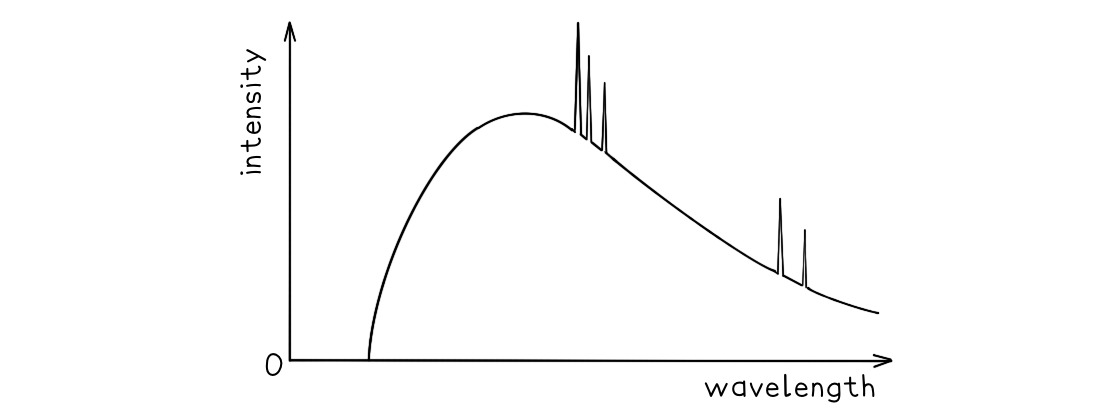 Explain why:
a) A continuous spectrum of wavelengths is produced.
b) The spectrum has a sharp cut-off at short wavelengths.
Part (a)
- Photons are produced whenever a charged particle is accelerated towards a metal target
- The wavelength of the photons depends on the magnitude of the acceleration
- The electrons which hit the target have a distribution of accelerations, therefore, a continuous spectrum of wavelengths is observed
Part (b)
- The minimum wavelength is equal to

- This equation shows the maximum energy of the electron corresponds to the minimum wavelength
- Therefore, the higher the acceleration, the shorter the wavelength
- At short wavelengths, the sharp cut-off occurs as each electron produces a single photon, so, all the electron energy is given up in one collision
- This equation shows the maximum energy of the electron corresponds to the minimum wavelength